The focus of our work is to examine the relationship between anatomical and biomechanical factors of the foot and ankle and mechanisms of injury, interventions, and treatment outcomes.
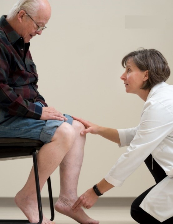
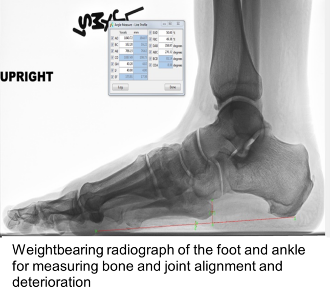
Our research combines multiple, non-invasive measures of muscle, tendon, and fascial function with kinematic analysis of movement. We have developed the methodology necessary to measure muscle, fat, tendon, and fascial volumes as well as bony alignment. The use of imaging techniques in physical therapy related research has allowed us to explore the contribution of tissue level abnormalities to foot and ankle dysfunction.

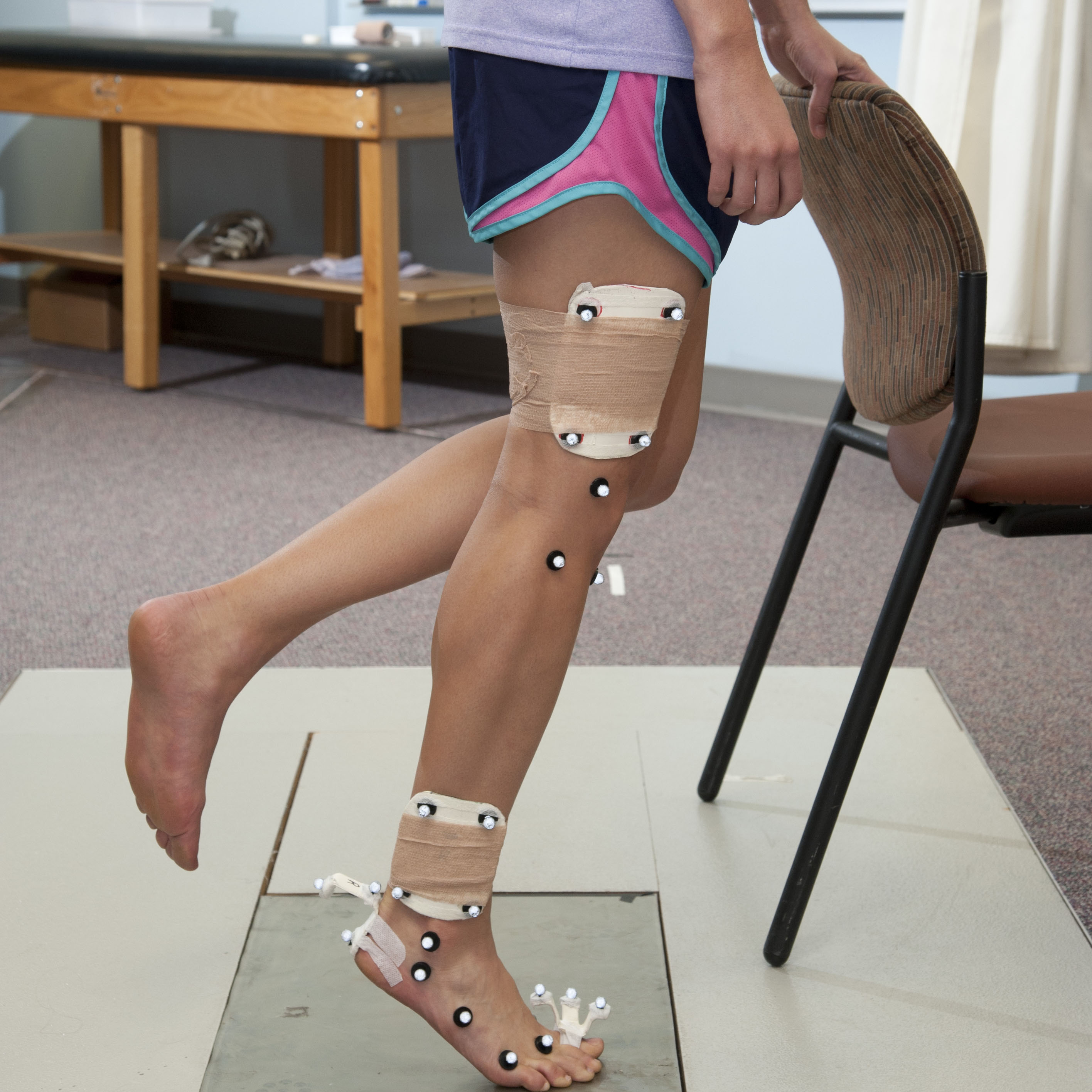
The kinematic analysis of foot and ankle motion is captured using a multi-segmental foot model. Previous work modeled the foot as a single segment with motion occurring at the ankle. The multi-segmental foot model allows measurement of motion within the foot. The expanded model allows us to discover the role of motion at the midfoot and toes during functional activities and to explore the contribution of the motion to foot deformity and pain.
Faculty Investigators
Mary K. Hastings, PT, DPT, MSCI, ATC [Profile ![]() ]
]
Student Members
Hyo-Jung Jeong, PT, MS, PhD, Postdoctoral Research Associate
Trenton Dickens, DPT Student
Current Research Studies
Chronic Kidney Disease-Mineral Bone Disorder (CKD-MBD) Syndrome in the diabetic, neuropathic foot.
Funding Source: NIH NIDDK 2R01 DK107809
Non-traumatic lower extremity amputations (NLEA) in people with diabetic peripheral neuropathy (DPN) have devastating consequences, most notable a 3-year mortality rate of up to 71%. The highest rate of NLEA occurs in individuals with diabetes-induced end-stage chronic kidney disease (CKD). CKD progression is marked by a bone-vascular paradox in which there is a loss of bone quantity & quality and an increase in vascular calcification, known as CKD-mineral bone disorder syndrome (CKD-MBD). Interestingly, our current research found DPN associated foot disease is also marked by a loss of BMD and vascular compromise. Yet, no one has explored the potential link between CKD-MBD, DPN foot disease, and the pathway to NLEA. Thus, the purpose this work is to examine DPN foot bone and vascular deterioration and test our hypothesis that CKD-MBD is a driving factor in the pathway to NLEA.
Muscle, joint, and movement deterioration contributing to neuropathic forefoot deformity
Funding Source: NIH/NIDDK R01 DK107809

Forefoot deformity in individuals with diabetes and neuropathy increases the risk of skin breakdown and possibility of lower extremity amputation. We will follow individuals with diabetes and neuropathy over 3 years and measure key factors we believe contribute to toe deformity (foot muscle and fat volumes, toe extension movement pattern, and advanced glycation end products). In addition we have included an intervention to assess the ability of a foot specific stretching and strengthening foot program to impact deformity progression.

The role of intermuscular fat in diabetic muscle pathology
Funding source: Academy of Foot and Ankle Society and Program in Physical Therapy, WashU Medicine
The pilot project is investigating the role of intermuscular adipose tissue in the lower extremity muscle pathology of individuals with diabetes. We are obtaining muscle and fat samples from the calf and foot and will use gene chip technology to understand how changes in IMAT-sensitive pathways might regulate muscle function.
Past Research Studies
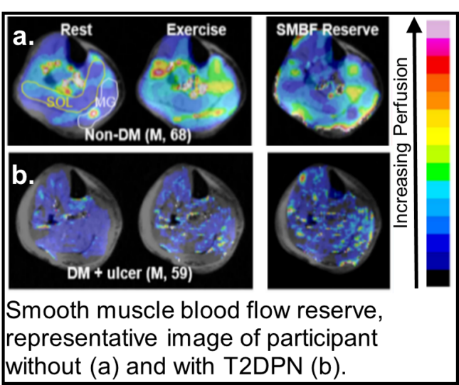
- Characterization of microcirculatory function in diabetic leg and foot with MRI: Individuals with diabetes have reduced ability to increase blood flow in skeletal muscle blood flow reserve is correlated with physical function.
- Biomechanical Contributions to Acquired Foot Deformity in Diabetes (Comprehensive Opportunities in Rehabilitation Research Training NIH/NICHD, NCMRR, NINDS K12 HD055931)
- Botulinum Toxin’s Effects on Plantar Ulcer Recurrence (NIH/NICH/NCMRR R21 HD048972)
- Biomarker development for Diabetic Complications (NIH/ NIDDK R21 DK079457)
- Outcomes of the Bridle procedure for the treatment of traumatic foot drop (Midwest Stone Institute)
- Effects of Discomfort and Lower Extremity Fatigue on Gait and Activity Parameters (Dr. Scholl’s)