We study patterns of synaptic communication within sensorimotor neural networks and how these networks learn to modify their function(s) when new patterns are introduced or existing patterns are altered. We are particularly interested in understanding the extent to which these basic principles of neural learning can be leveraged to restore a more appropriate balance of sensory and motor transmission after CNS injury.

Much of our work is motivated by the observation that injuries to the central nervous system frequently result both in movement impairments and changes in pain perception. Yet, while many therapies intended to target movement impairments also impact pain perception (and vice versa), these interventions are developed and evaluated primarily through the prism of the movement or pain alone.
Our work is predicated on the notion that optimal therapies for restoring function after neurological injury must be grounded in a neuromechanistic understanding of the causes of impairment, which requires an integrative view of nervous system function and an interdisciplinary approach to research. As a result, our work is translational in nature, spanning basic science, pre-clinical (animal), and clinical (human-subjects) neurophysiology and neurological rehabilitation research in the context of normative physiology, spinal cord injury, and stroke.
The ultimate goal of our work is to reduce maladaptive consequences of altered sensorimotor communication (e.g., neuropathic pain, movement impairments) after CNS injury towards development of therapies that offer multi-modal rehabilitation.
For more information, please see Dr. Jacob McPherson’s laboratory website: https://sites.wustl.edu/pmrflab
Faculty Investigator

Lab Members
Jeremie Ferey, PhD, Lab
Manager

Gerson Romero Moreno,
PhD Student, Biomedical
Engineering

Avery Twyman, PhD Candidate,
Biomedical Engineering

Jane Wu, Undergraduate Research
Assistant

Lucia Lopez, MS, Research
Assistant
Current Research Studies
Intraspinal functional connectivity and neural plasticity (basic science, pre-clinical)
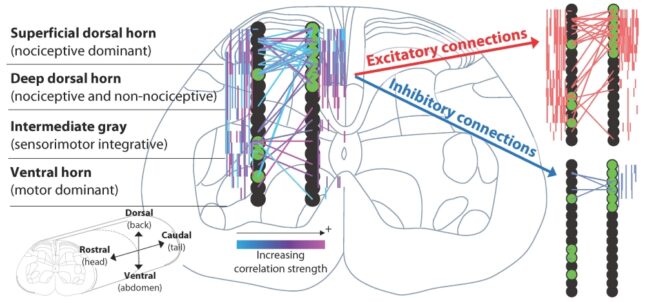
Perception and action are central to our ability to interact with and learn from the world around us. Yet, we are only beginning to appreciate how the nervous system makes staggeringly complex tasks and decisions seem routine. In this line of research, we study the spinal cord’s role in these processes. The spinal cord is the first stop in the CNS for sensory feedback coming from the body (perception) and the last stop in the CNS for sending motor commands out to muscles (action). Therefore, it presents a unique opportunity for studying communication within sensory and motor networks and how these networks learn and adapt.
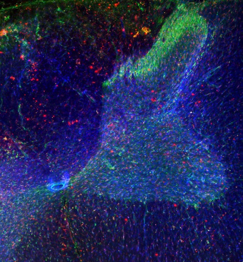
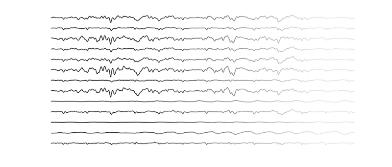
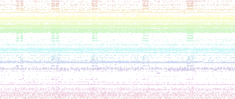
Currently, we are studying whether seemingly spontaneous patterns of neural activity in spinal networks may actually represent (at least in part) a ‘memory’ or ‘replay’ of past experiences and a default state of readiness to execute behaviors – even when unconscious. We are also studying intraspinal local field potentials, a type of biophysical signal that reflects the aggregate neural activity of a large population of simultaneously active neurons. Specifically, we want to understand whether these signals convey meaningful information between functionally and spatially different neural structures, in effect representing a different language that networks of neurons could use to communicate (in addition to direct synaptic information transfer from one neuron to another). These studies are primarily conducted in vivo in rats using electrophysiological recordings of hundreds to thousands of neurons in the spinal cord, as well as recordings from peripheral nerves and muscles.
Intraspinal microstimulation for multi-modal rehabilitation (basic science, pre-clinical)
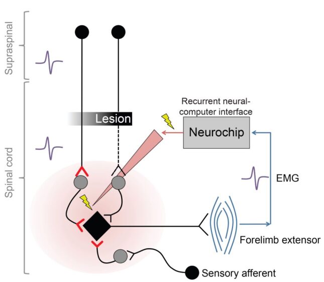
Spinal cord injury (SCI) often results in motor impairments and neuropathic pain. These conditions are related to changes in neural transmission in regions of the spinal cord that control motor output and sensory processing. Generally, there is too little neural transmission in spinal motor pathways below the lesion, whereas there is excessive, inappropriate neural transmission in pain pathways below the lesion.
We have previously developed a recurrent neural computer interface that allows us to deliver small amounts of electrical current inside the spinal cord within a few milliseconds of detecting specific, functionally related neural activity in other regions. This so-called ‘closed loop’ intraspinal microstimulation approach allows us to restore natural patterns of neural transmission in sensorimotor pathways that span an SCI by artificially connecting neurons that can still be activated voluntarily with those that are weakened or cannot be activated voluntarily due to the lesion. Once the appropriate patterns of neural activity have been (re)introduced, they can be reinforced over time by taking advantage of a fundamental type of neural learning called spike-timing-dependent plasticity, in which the strength of neural connections can be increased or decreased when one neuron repeatedly contributes to the firing of another. We have shown that this approach can lead to long-term improvements in elbow, wrist, and digit control in chronic cervical SCI.
Currently, we are exploring the extent to which intraspinal microstimulation for motor rehabilitation can also be designed to reduce pathologically increased transmission in spinal pain pathways below an SCI. These studies are conducted in vivo in rats using a combination of neural-computer interfaces, targeted neuropharmacology, and physical rehabilitation.
Intraspinal microstimulation of the ventral horn has therapeutically relevant cross-modal effects on nociception. Brain Communications.
Unintentionally intentional: unintended effects of spinal stimulation as a platform for multimodal neurorehabilitation after spinal cord injury. Bioelectronic Medicine.
Spinal stimulation for motor rehabilitation immediately modulates nociceptive transmission. J Neural Engineering.
Rehabilitative neuroplasticity in brainstem-spinal neural pathways (pre-clinical, clinical)
If a person is trying to escape from a frightening or stressful situation, like running away from a bear while on a hike, they will likely find that they’re able to run much faster than when they’re just on a daily jog. And if they stub their toe along the way, they probably won’t notice the pain. On the other hand, if a person is falling asleep in class after a long night and was asked to do as many pushups as possible, they’d likely not be at peak performance. But their toe would absolutely kill when they stood up and stubbed it on their desk.
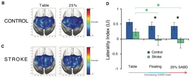
You can thank a region of the brainstem known as the ponto-medullary reticular formation (PMRF) for both of these phenomena. Neural pathways descending from the PMRF to the spinal cord release a type of neurotransmitter that dramatically increases motor-related neural transmission while it simultaneously reduces pain-related transmission. Our research suggests that neural transmission from the PMRF to the spinal cord also becomes pathologically increased after a stroke, and appears to be a key contributor to many common post-stroke movement impairments.
Currently, we are studying interrelationships between the neural control of movement and pain perception to further our understanding of how altered transmission from the PMRF to the spinal cord contributes to movement and sensory impairments. We are also attempting to reduce pathologically increased transmission in these same pathways by using non-invasive electrical stimulation to drive restorative neuroplasticity. These studies are conducted in individuals with and without chronic hemiparetic stroke using a combination of non-invasive electrical stimulation, mechatronic devices/robotics, electromyography, and targeted neuropharmacology.